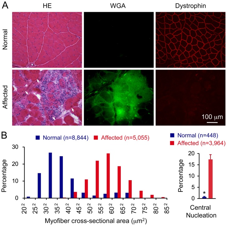
Figure 1. Dystrophin deficiency leads to severe muscle damage in the extensor carpi ulnaris muscle in affected dogs. A,
Haematoxylin and eosin (HE) staining (left panel), wheat germ agglutinin (WGA) staining (middle panel) and dystrophin immunofluoresence staining (right panel) of the extensor carpi ulnaris muscle from a normal dog (top panel) and an affected dog (bottom panel). WGA staining reveals tissue fibrosis. Arrow, regenerative myofibers with centrally located nuclei. B, Morphometric quantification of the myofiber size (left panel) and central nucleation in normal and affected ECU muscles. The myofiber size is expressed according to the distribution of the cross-sectional area of individual myofibers. Results for the normal ECU muscle was from 8,844 myofibers of five normal dogs. Results for the dystrophic ECU muscle was from 5,055 myofibers of four affected dogs. The central nucleation is expressed as the percentage of myofiber with centrally localized nuclei among all myofibers counted. Results for the normal ECU muscle was from 9,467 myofibers of five normal dogs. Results for the dystrophic ECU muscle was from 3,964 myofibers of four affected dogs. Asterisk, significantly different (p = 0.0001).

