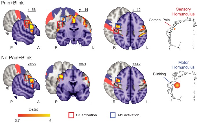
Figure 1. Somatotopic activation triggered by combined corneal pain and eye blink.
The Pain+Blink condition activated contralateral S1 (Max zstat = 4.9 at 56, −14, 43) and bilateral M1 (Max zstat = 9.9 at 52, 2, 31) in regions corresponding to the eye in the Penfield sensory and motor homunculi [19] (p<0.0001, uncorrected for multiple comparisons). The No Pain+Blink condition activated bilateral M1 (Max zstat = 7.4 at 44, −4, 43), but not S1. Investigations were restricted to the non-shaded areas in the activation maps, which correspond to bilateral pre- (blue) and post-central gyri (red) as highlighted in the underlying brain slices and colored squares (dashed squares denote absent activation). Note that the boundaries of these probabilistic-defined areas overlap with other regions, such as supplementary motor area, middle frontal gyrus, and supramarginal gyrus. Of note, the supplementary motor area has previously been associated with voluntary blinking [13], and was active in both conditions along the midline of coronal slice y = −14 (only shown for Pain+Blink in figure). A = anterior; L = left; P = posterior; R = right.

