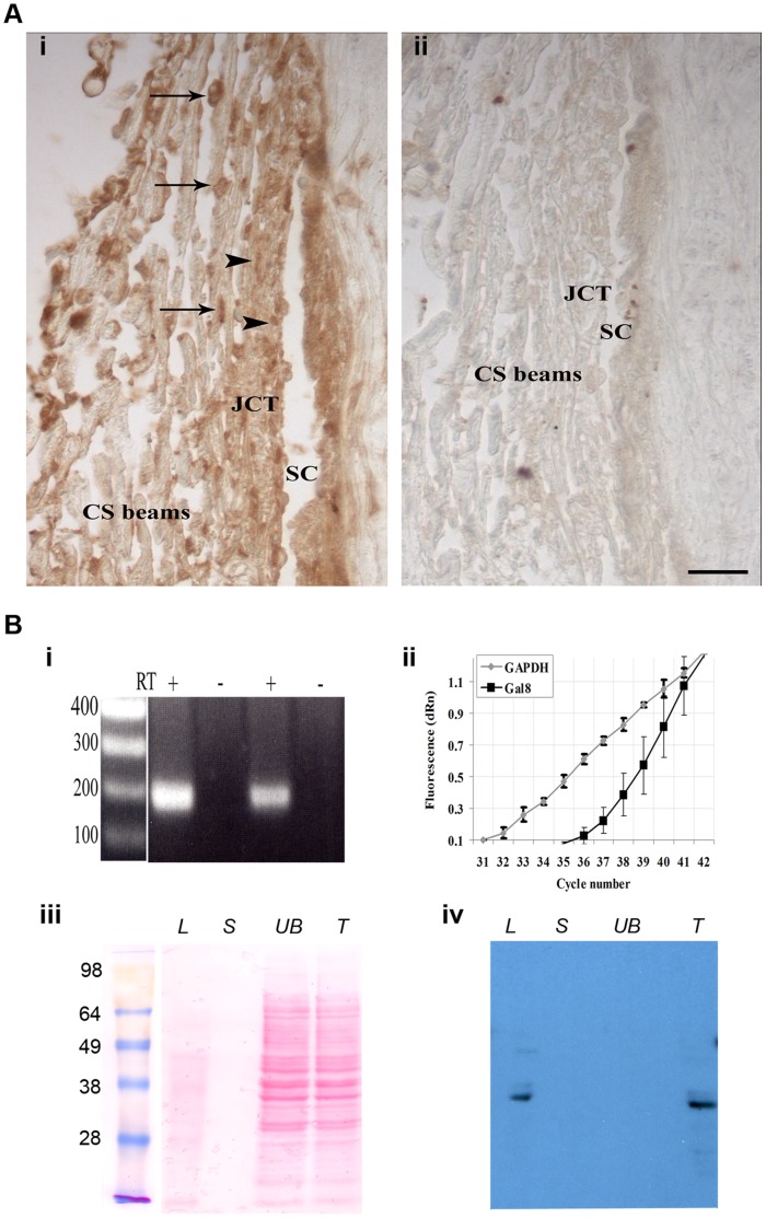
Figure 1. Galectin-8 is expressed in the Trabecular Meshwork (TM).
A: paraffin sections of anterior chamber angle from a normal human eye were immunostained with anti-Gal8 antibody. (i) anti-Gal8 IgG reacted intensely with cells on the trabecular beams (arrows) and with cells in the juxtacanalicular portion of TM (arrowheads). Staining was also observed in the ECM of both portions of TM (JCT and CS) and in the wall of Schlemm’s canal. (ii) No staining was observed when the sections were not exposed to the primary antibody. SC: Schlemm’s canal, JCT: juxtacanalicular TM; CS beams: corneoscleral beams. Bar: 25 µm. B: (i) RT-PCR. Total RNA (1.0 µg) from confluent cultures of normal human TM cells was subjected to RT-PCR. The expected 191 bp fragment was amplified using Gal8- specific-primers. In each case, no components were amplified when reaction mixtures lacked reverse transcriptase (RT). (ii) qRT-PCR. Total RNA was subjected to Taq-Man RT-PCR using Gal8 specific primers. Original amplification plots of Gal8 and GAPDH mRNAs genes are shown (Ct 37.37 and 33.38 for Gal8 and GAPDH, respectively). N = 3 for each experiment; all experiments were performed twice using TM cells from two different donors with reproducible results. (iii and iv) Western Blot Analysis. Protein extracts from confluent cultures of normal human TM cells were incubated with lactogel beads and eluted first with sucrose, and then with lactose. Eluted proteins were electrophoresed, the protein blot of the gel was stained with Ponceau S (iii) and was then processed for immunostaining with goat anti-Gal8 (iv). Both the total cell extract (T) and the lactose eluate (L) contained a major 36-kDa anti-Gal8 reactive component. This component was not detected in the unbound fraction (UB) and in the sucrose eluate (S).

