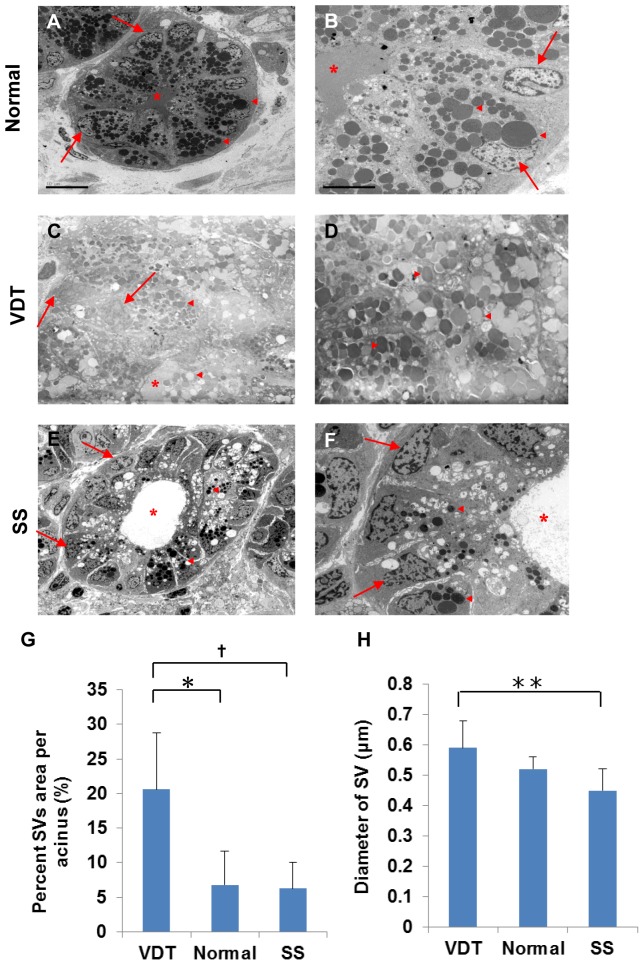
Figure 3. Electron microscopic findings of the lacrimal gland acinus in the three groups.
A: SV accumulation in the normal lacrimal gland (n = 4). Scale bars = 10 µm. B: Homogeneous SVs in normal controls. Scale bars = 5 µm. C: Excessive accumulation of SVs in the VDT group (n = 4). D: High magnification view of SVs in the VDT group. E: Only a few SVs in the SS group (n = 12). F: High magnification view of SVs in the SS group. G: Percent SV area per acinus. Total number of acini examined/group: 20/VDT group (4 cases), 58/SS group (12 cases), and 20/Normal controls (4 cases). *P = 0.021, † P = 0.004 (Mann-Whitney U test). H: SV diameter. Total number of vesicles examined/group: 6274/VDT group (4 cases), 11837/SS group (12 cases), and 2875/Normal controls (4 cases). **P = 0.025 (Mann-Whitney U test). Original magnification: ×2000 (A, C, E), ×5000 (B, D, F). Asterisk, Ductal lumen; Arrows, Nuclei; Triangle, SV.

