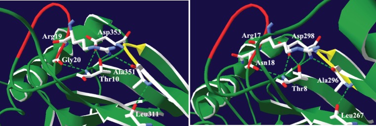
Figure 8. Depiction of the predicted hydrogen bonding network in the AxDx strand/loop region of human and B. subtilus tRNase ZSs.
The atomic coordinates of human and B. subtilus tRNase ZSs were obtained from the Protein Data Bank, www.rcsb.org (PDB accession codes 3ZWF and 1Y44, respectively). Images were made with Swiss-PdbViewer [59]. Potential hydrogen bonds were determined using the Swiss-PdbViewer and Insight II. Secondary structure elements are colored green. The AxDx strand/loop is colored yellow and the PxPxRG or PxKxRN loop is colored red. Hydrogen bonds are represented by a dashed green line. (A) In the human tRNase ZS, the AxDx strand/loop-centered hydrogen bond network involves Ala351 (O) and Arg19 (NH1), Asp353 (OD2) and Gly20 (HN), Ala351 (HN) and Leu311 (O), Asp353 (OD1) and Thr10 (HN and OG1), and Gly20 (O) and Thr10 (HN). (B) In B. subtilus, the AxDx strand/loop-centered hydrogen bond network includes Ala296 (O) and Arg17 (NH1), Asp298 (OD2) and Asn18 (HN), Ala 296 (HN) and Leu267 (O), Asp298 (OD2) and Thr8 (HN and OG1), and Asn18 (O) and Thr8 (HN).

