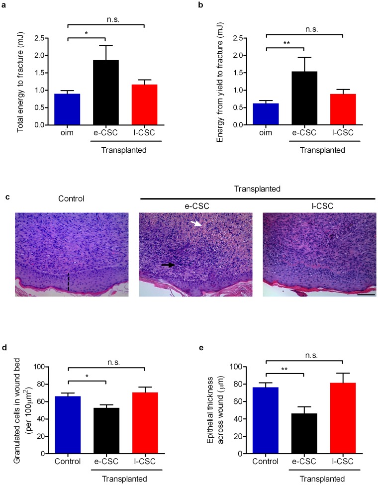
Figure 9. e-CSC showed better tissue repair capacity in vivo compared to l-CSC.
(a) Mechanical 3-point bending data in a mouse model of bone brittleness (oim) for overall bone quality shown by total energy input in millijoules to fracture and for (b) femoral plasticity shown by energy input in millijoules from yield to fracture. Results shown per mouse for non-transplanted oim (blue) and oim transplanted with e-CSC (black) and l-CSC (red). (c) Cross-section of skin wounds 7 days after 4 mm dermal biopsy in mice treated with either PBS alone (Control) or transplanted with e-CSC or l-CSC; stained with haematoxylin and eosin. Indicated is collagen (white arrow), granulated cells (black arrow) and epithelial thickness (dashed line). In the same model of wound repair, (d) number of granulated cells in the wound bed (per 100 µm2) and (e) epithelial thickness across wound in µm. Data. * P<0.05; ** P<0.01, n.s. (not significant), Student's t test. Mean ± s.e.m.

