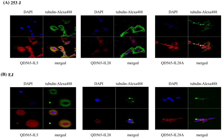
Figure 8. Confocal microscopy analysis of IL-5, IL-20, and IL-28A in bladder cancer 253J and EJ cells.
(A, B) 253J and EJ cells, respectively, were stained with antibodies against QD565-conjugated IL-5, IL-20, and IL-28A (red). Both nuclei (anti-DAPI) and cytoplasm (anti-tubulin-Alexa488) are counterstained with DAPI (blue) and tubulin-Alexa488 (green).

