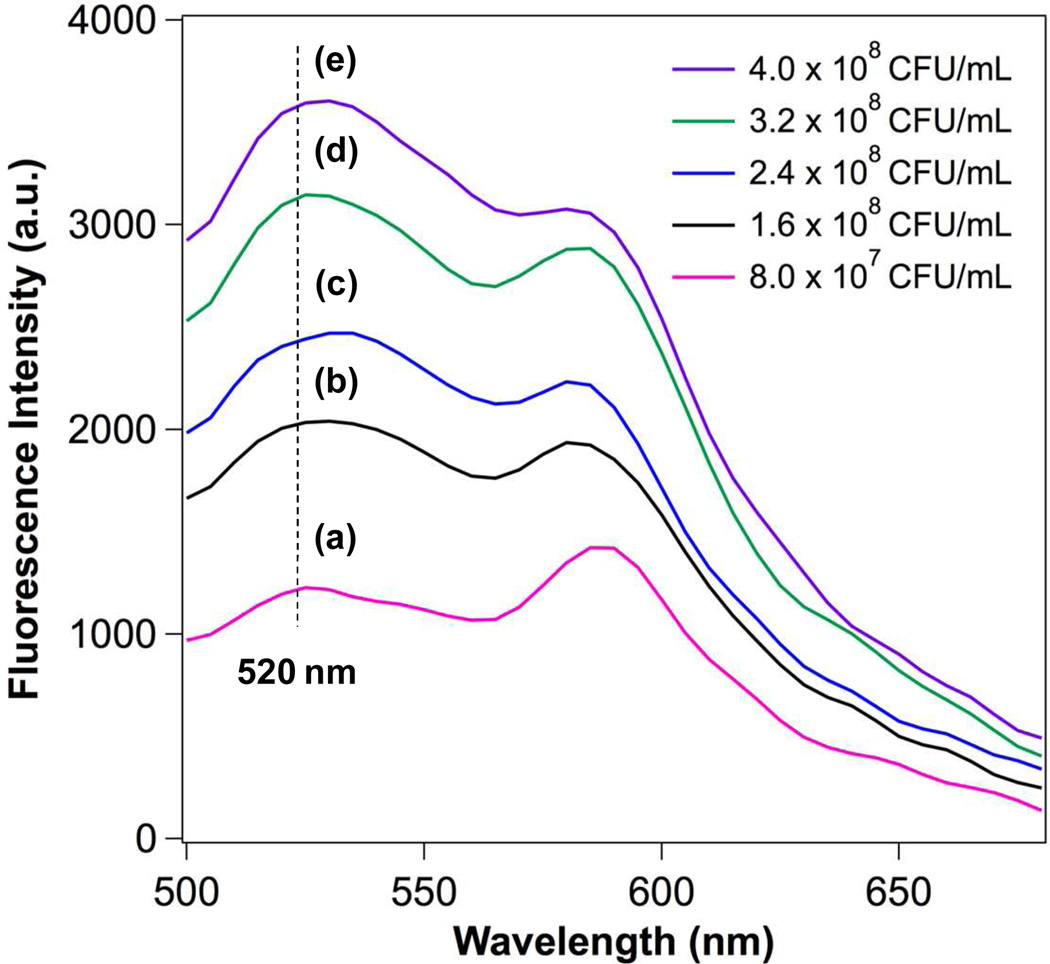
Figure 3.
FRET measurements of the fusion between LipoLLA and H. pylori SS1. A fluorescent donor (C6NBD, 0.1 mol%) and a fluorescent acceptor (DMPE-RhB, 0.5 mol%) were concurrently incorporated into the lipid bilayer membranes of LipoLLA so that the acceptor completely quenched the fluorescence emission from the donor. The FRET-pair labeled LipoLLA was incubated with H. pylori at a concentration of a–e: 8.0×10⁷, 1.6×10⁸, 2.4×10⁸, 3.2×10⁸, and 4×10⁸ CFU/mL for 10 min. After removing the excess LipoLLA, all samples were excited at 470 nm. A rise in emission intensity of C6NBD (donor) at 520 nm was observed with the increase of bacterial concentrations, indicating the occurrence of fusion between LipoLLA and H. pylori that caused the spatial separation of C6NBD and DMPE-RhB.

