
Figure 1.
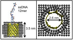
(Left) Simulation setup. Protein β-barrel is shown in ribbon, cut-away format. The membrane-mimetic slab is shown as spheres. The DNA strand is shown in licorice format. (Right) Pore dimensions, as viewed down the barrel principle axis.
Official websites use .gov
A
.gov website belongs to an official
government organization in the United States.
Secure .gov websites use HTTPS
A lock (
) or https:// means you've safely
connected to the .gov website. Share sensitive
information only on official, secure websites.
(Left) Simulation setup. Protein β-barrel is shown in ribbon, cut-away format. The membrane-mimetic slab is shown as spheres. The DNA strand is shown in licorice format. (Right) Pore dimensions, as viewed down the barrel principle axis.