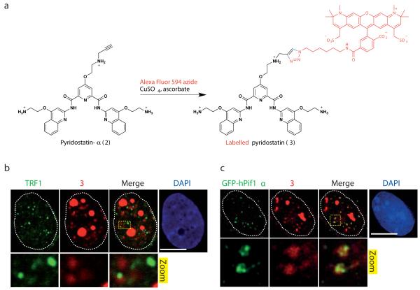
Figure 4. Pyridostatin and hPif1 targeted overlapping sites in cells.
(a) Molecular structure of 2 and synthetic scheme for generating 3 in cells; a single isomer is shown for clarity, Alexa Fluor 594 is marked in red and newly formed chemical bonds are marked in blue. (b) 3 formed nuclear foci mainly at non-telomeric sites in MRC5-SV40 cells, fixed with formaldehyde prior to incubation with 2 followed by chemical labelling; dotted white lines indicate nuclear peripheries; zoomed images correspond to further 4X magnification. (c) GFP-hPif1α expressing U2OS cells display small nuclear foci of GFP-hPif1α that co-localizes with 3 in cells fixed with formaldehyde prior to incubation with 2 followed by chemical labelling; dotted white lines indicate nuclear peripheries; zoomed images correspond to further 5X magnification. Note that cells were first pre-extracted with CSK buffer as described in Supplementary Methods, then fixed with formaldehyde and stained with the indicated antibody. Scale bar, 10 μm.

