Figure 1.
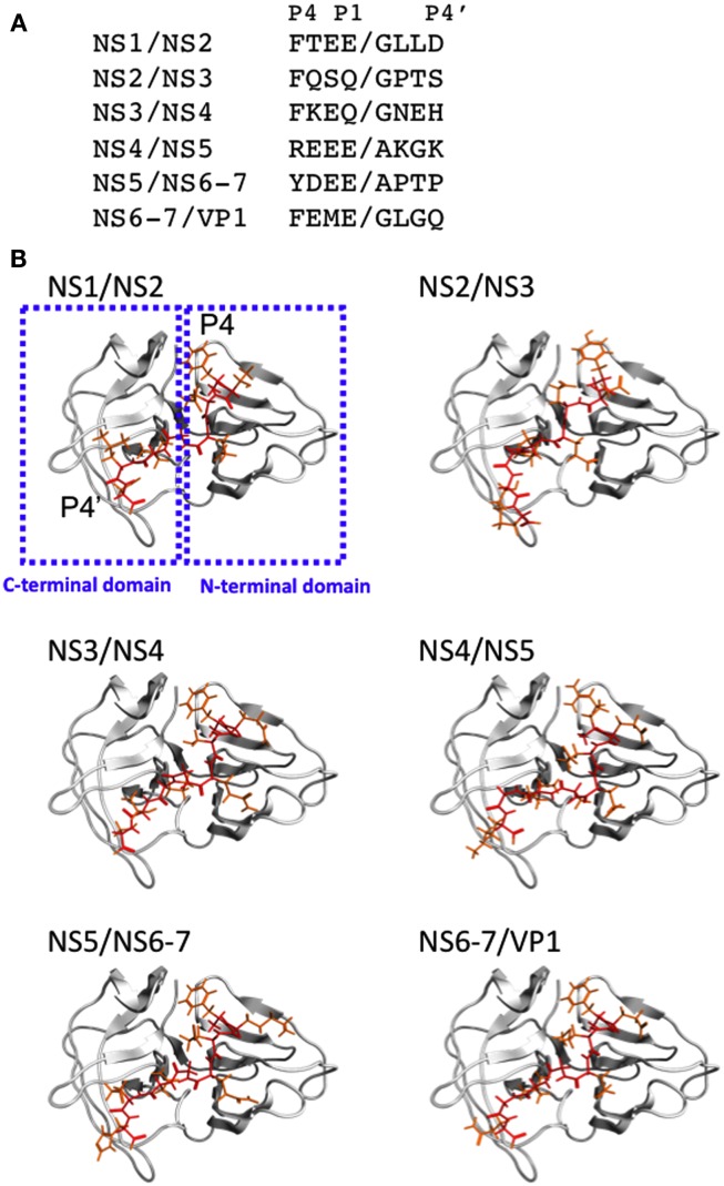
Structural models of SaV protease docked to the substrate octapeptides. (A) Sequences of the six cleavage sites of the SaV ORF1 polyprotein are shown with one-letter amino acid codes. Slashes represent the peptide bonds cleaved by the protease. (B) Structural models of the SaV protease-substrate complex. The 3-D structural model of the protease domain was constructed by homology modeling and thermodynamically and physically refined as described previously (Oka et al., 2007). The 3-D structural models of the octapeptides corresponding to the six authentic cleavage sites of the SaV Mc10 ORF1 were constructed by using the Molecular Builder tool in MOE. The optimized protease model was docked to individual octapeptides using the automated ligand docking program ASEDock2005 (Goto et al., 2008) operated in MOE as described previously (Yokoyama et al., 2010). Red and orange sticks indicate main and side chains of the octapeptides, respectively.

