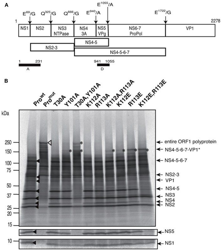
Figure 4.
Site-directed mutagenesis of the substrate interaction sites of SaV Mc10 protease. (A) Proteolytic cleavage map of the SaV Mc10 ORF1 polyprotein and the processing intermediates (Oka et al., 2006). Black bars indicate the protein segments, A and D, used to raise polyclonal antibodies for detection of the NS1 and NS5 proteins, respectively. (B) SDS-PAGE of 35S-labeled in vitro translation products of SaV Mc10 ORF1 containing various protease mutants. NS1 and NS5 were detected by immunoprecipitation using anti-A or anti-D polyclonal antibodies as described previously (Oka et al., 2005b, 2006, 2009). Mc10 ORF1 containing functional protease (Prowt) and a defective mutant lacking in the proteolysis activity (Promut) were included as described previously (Oka et al., 2005b). Newly appearing products when compared to Prowt are indicated by asterisks. Size markers are shown on the left. Mc10 ORF1-specific proteins (Oka et al., 2005b, 2006) are shown on the right.

