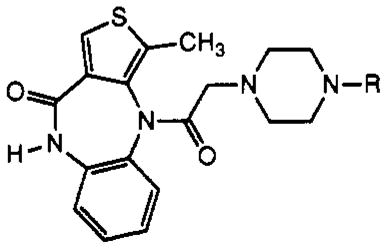
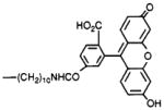
Table I.
Structures of Telenzepine Derivatives Synthesized and Their Affinity at Muscarinic Receptors

| ||||
|---|---|---|---|---|
| no. | R | receptor subtype and Ki valuesa (nM)
|
||
| forebrain | heart | other | ||
| 1 | —CH3 (telenzepine) | 4.67 ± 0.53 | 66.9 ± 32 | m1 (A9L), 1.8; m3, 6.9; m4,17.4 |
| 2 | —H | b | b | |
| 4 | —(CH2)10NH2 | b | 3.7 | m1 (A9L), 2.39; m3, 7.5; m4, 1.3 |
| 5 |
|
2.22 ± 0.48 | 1.75 ± 0.52 | |
| 6 |
|
0.29 ± 0.03 | 0.31 ± 0.07 | |
| 7 |
|
b | b | |
| 8 |
|
1.25 ± 0.12 | 3.7 ± 0.5 | |
| 9 |

|
3.20 ± 1.33 | 0.60 ± 0.22 | |
| 10 |
|
42.5 ± 10.0 | 43.7 ± 12.2 | |
| 11 |

|
10.5 ± 0.7 | b | |
| 12 |

|
116 ± 17.5 | 82.1 ± 11 | |
| 13 |
|
1.21 ± 0.12 | 1.38 ± 0.15 | |
| 14 |

|
20.1 ± 6.2 | b | |
| 15 |

|
11.8 ± 3.4 | b | |
Ki values for inhibition of binding of [3H]NMS are given. Values are expressed as average for a single experiment run in triplicate or as mean ± SEM for three experiments. Membranes from rat brain (mostly m1-receptors) or rat heart (m2-receptors) were isolated as described in Experimental Procedures. Varying concentrations of compound were incubated with 0.5 nM [3H]NMS and a quantity of membranes containing 100–300 μg of protein for 60 min at 37 °C and then rapidly filtered over GF/B filters. Competition binding experiments using membranes from transfected A9L cells (m1- and m3-receptors) and from NG108–15 cells (m4-receptors) were carried out as previously described (14). The resulting data were analyzed by nonlinear regression, and IC50 values were converted to Ki values using the Cheng–Prusoff equation (15) with the following Kd values for [3H]NMS binding: 0.350 nM with brain membranes and 0.425 nM with cardiac membranes.
Not determined.
