Fig. 1.
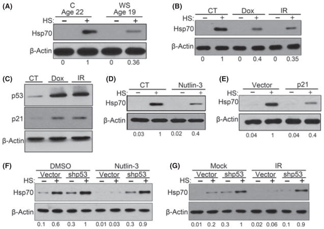
Activation of p53 pathway caused suppression of HSR. (A) WS fibroblasts (passage 4) and age-matched C fibroblasts (passage 15) were heat shocked and were collected for immunoblotting. (B) TIG-1 fibroblasts were treated with 100 nM of Dox overnight or 10 Gy IR. After 6 days, cells were heat shocked and incubated for 6 h before collection. (C) Same cells from (B) were immunoblotted for indicated proteins. (D) Cells that were induced senescent by 5 days of 10 μM nutlin-3 treatment were heat shocked, and Hsp70 levels were measured. (E) Hsp70 accumulation after heat shock in cells that overexpress p21. (F) TIG-1 cells were infected with empty or shp53 retroviruses, and nutlin-3 was added for additional 5 days. Cells were collected 6 h after heat shock and immunoblotted for Hsp70. Interestingly, we observed an increase in basal level and after heat shock level of Hsp70 upon p53 depletion. (G) Control or p53-depleted cells were treated with 6 Gy IR and cultured for 5 days. Cells were collected 6 h after heat shock and immunoblotted for Hsp70. Abbreviations: C, age-matched healthy control; CT, control.

