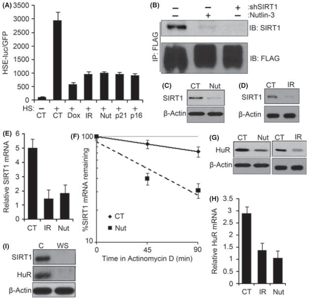
Fig. 3.
DNA-damage-induced senescence suppresses Hsf1 activity by decreasing SIRT1. (A) TIG-1 fibroblasts were infected with the HSE-luciferase and treated with 100 nM Dox overnight and cultured for 6 days; 10 Gy IR and cultured for 6 days; 10 μM nutlin-3 for 5 days or retroviral expression of p21 or p16 for 6 days. After heat shock, cells were incubated for 6 h before collection, and luciferase activity was measured. The means and ±SEM indicate three independent experiments. (B) FLAG-tagged wild-type Hsf1 was expressed with retrovirus in TIG-1 fibroblasts and treated with 10 μM of nutlin-3 for 5 days to induce senescence or depleted of SIRT1 by lentiviral shRNA. Cells were collected promptly after heat shock at 43 °C for 1 h, and immunoprecipitated with anti-FLAG antibody. The precipitates were immunoblotted for SIRT1. (C and D) Early passage TIG-1 fibroblasts were treated as in (A) and immunoblotted for SIRT1. (E) TIG-1 fibroblasts exposed to conditions mentioned (A) were collected for RNA, and qRT-PCR was performed using SIRT1 and GAPDH (housekeeping gene for control) mRNA-specific primers. The means and ±SEM are from three independent experiments. (F) SIRT1 mRNA half-life was measured in control (CT) and nutlin-3-induced senescent cells (Nut) by incubating with 5 μg mL−1 of actinomycin D and collecting RNA after 45 and 90 min. SIRT1 mRNA was normalized for GAPDH mRNA. The means and ±SEM were calculated from triplicates of two independent experiments. (G) Early passage TIG-1 fibroblasts were exposed to conditions mentioned in (A), and HuR protein levels were measured by immunoblotting. (H) HuR mRNA levels were measured by qRT-PCR, as in (F). The means and ±SEM are from three independent experiments. (I) Same set of cells as in Fig. 1(A) were immunoblotted for SIRT1 and HuR.

