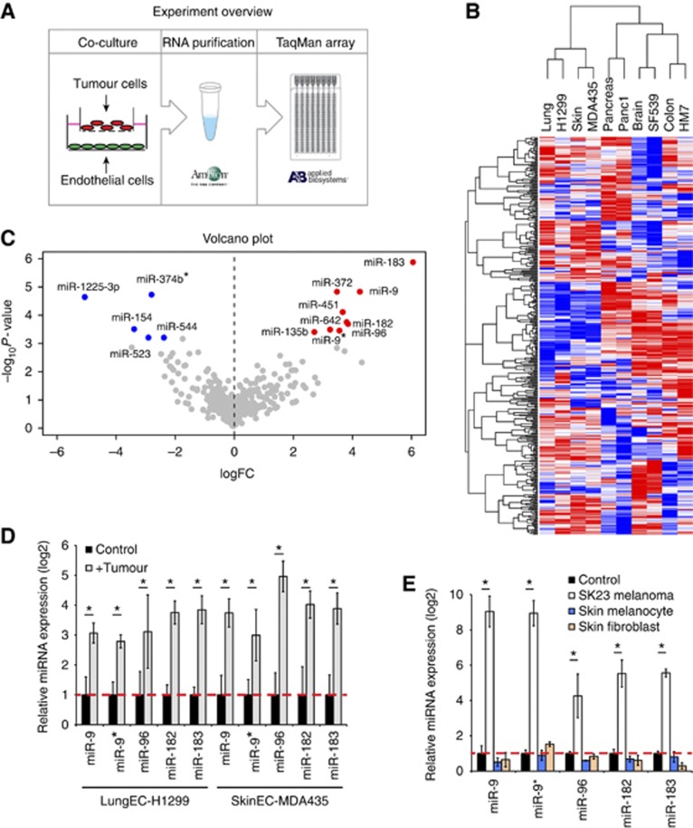
Figure 1.
Tumour cells induce upregulation of miRNAs in endothelial cells. (A) Experimental overview. (B) Hierarchical clustering of miRNAs in control or tumour-stimulated endothelial cells. (C) Volcano plot of miRNA changes across five pairs of tumour-endothelial cells. (D) Verification of tumour-induced miRNAs by quantitative RT–PCR. Lung endothelial cells co-cultured with H1299 and skin endothelial cells co-cultured with MDA435 for 24 h are shown. The experiment was repeated three times. *P<0.05, Student’s t-test. (E) Quantitative PCR of miRNAs in skin microvesicular endothelial cells stimulated with SK23 melanoma cells, normal skin melanocytes or fibroblasts. The experiment was repeated twice. *P<0.05, Student’s t-test.

