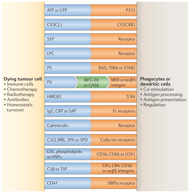
Figure 4. DC interaction with tumour cells: antigen capture.
The figure illustrates some phagocyte surface receptors and their putative ligands that are implicated in the recognition of dying tumour cells. Receptors shown in blue represent molecules expressed by dying tumour cells, receptors shown in orange represent molecules expressed by dendritic cells (DCs) and the receptors shown in green function as a ‘bridge’ between the two cells. BAI1, brain-specific angiogenesis inhibitor 1; C1Q, complement C1q subcomponent; C3β, complement C3β; CRP, cysteine-rich protein; GAS6, growth arrest-specific protein 6; HMGB1, high mobility group protein B1; HSP, heat shock protein; LDL, low-density lipoprotein; LOX1, lectin-like oxidized LDL receptor 1; LPC, lipid lysophosphatidylcholine; MBL, mannose binding lectin; MFG-E8, milk fat globule-EGF factor 8 (also known as lactadherin); P2Y2, P2Y purinoceptor 2; PS, phosphatidylserine; S1P, sphingosine 1-phosphate; SAP, sphingolipid activator protein 1; SIRPα, signal-regulatory protein-α; STAB2, stabilin 2; TIM4, T cell membrane protein 4; TLR4, Toll-like receptor 4; TSP, thrombospondin.

