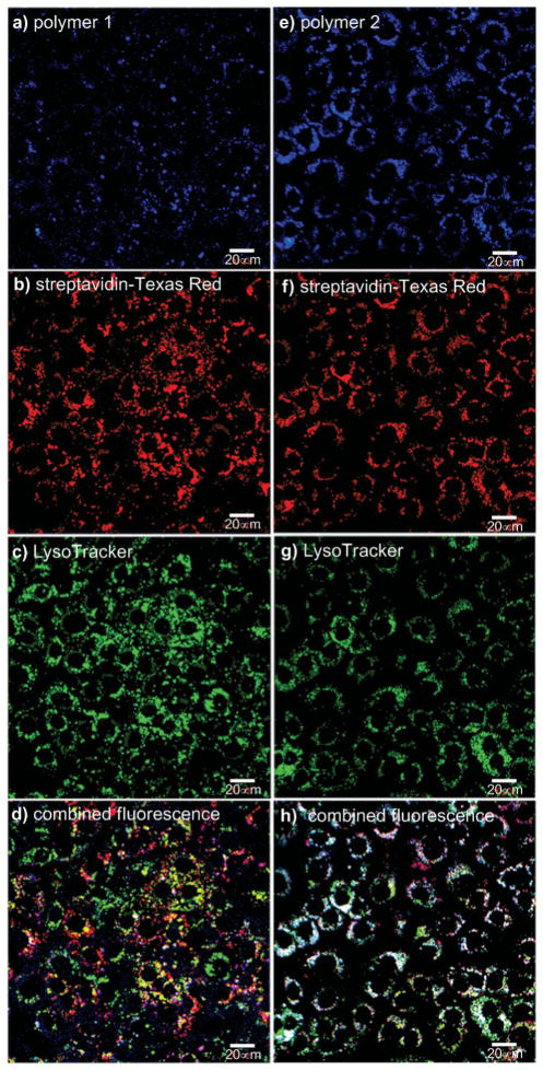
Fig. 3.
Delivery of streptavidin-Texas Red® into Clone 9 cells at 37 °C mediated by 1 and 2. Data for transport using polymer 1: (a) polymer 1 channel; (b) streptavidin-Texas Red® channel; (c) LysoTracker® Green; (d) colocalization shows mostly distinct blue, red, and green areas. Data for transport using polymer 2: (e) polymer 2 channel; (f) streptavidin-Texas Red® channel; (g) LysoTracker® Green; (h) colocalization shows mainly white areas where all three labels coexist. Throughout, the carrier (1.0 μM), streptavidin-Texas Red® (1.0 μM), and the Clone 9 cells were incubated at 37 °C for 15 h; the cells were then washed 3× with PBS buffer and 3× with heparin and analysed via fluorescence microscopy. Scale bar is 20 μm.

