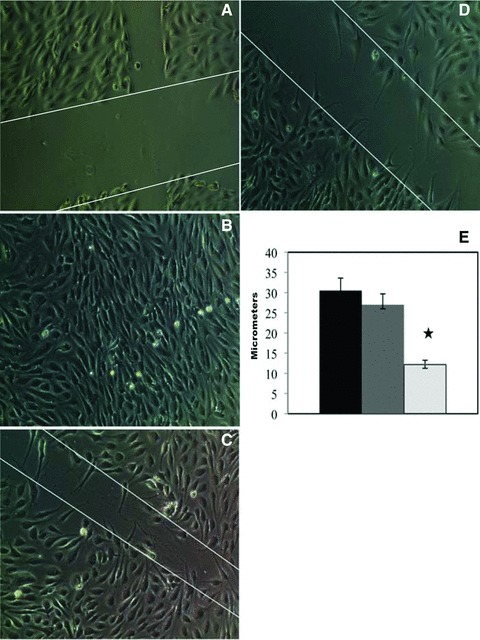
Fig 1.

Effects of basal and mobilized PBMCs on endothelial proliferation and migration. Wounded ECs were cultured under medium without (A) and with (B) endothelial growth factor supplements for 24 hrs, as negative and positive controls. When mobilized PBMNCs were co-cultured for 24 hrs with the wounded EC layer in the absence of endothelial growth factors, wound healing was accelerated as evidenced by more rapid endothelial proliferation and migration (C) by comparison with basal PBMNCs (D). Significant differences (P < 0.05) existed in the width of the wound after the healing process for wounds treated with mobilized cells (light grey) versus basal cells (dark grey) versus negative control (black) as shown in (E). Original magnification, χ200.
