
Figure 5.
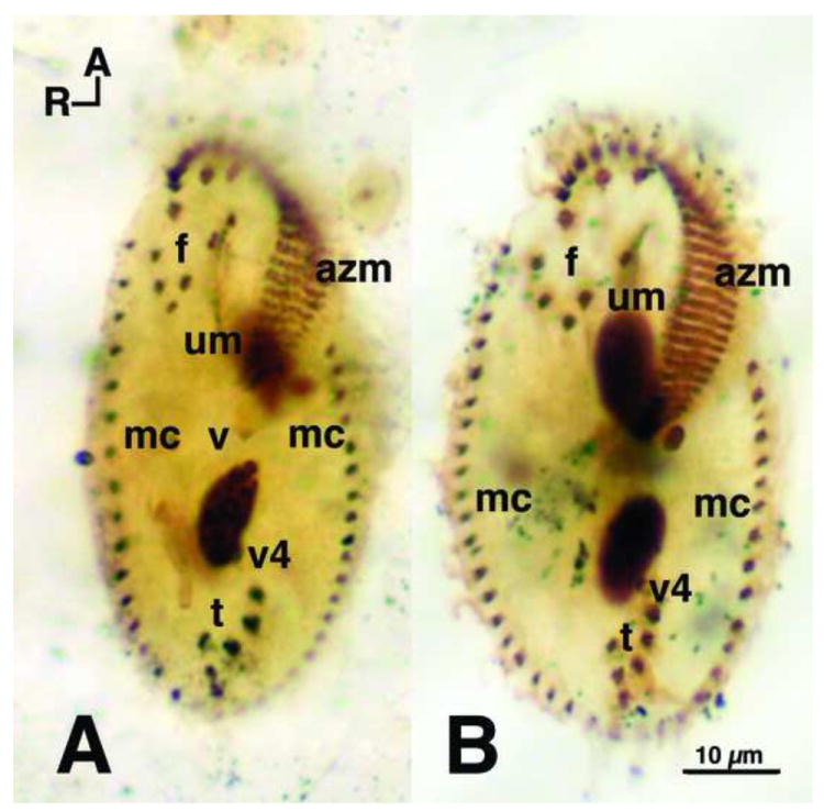
Protargol comparison of O. fallax and O. trifallax. Protargol stained cells, fixed with Perenyi’s fixative, showing the classic Oxytricha ventral surface ciliature. A. Oxytricha fallax stock 9D1 (the original Grimes stock) and B. Oxytricha trifallax JRB310, both showing the standard (Berger 1999) 18 fronto-ventral-transverse (fvt) cirri with 8 frontal cirri (f), 4 ventral cirri (v) (not all 4 shown because of focal level), and 6 transverse (t) cirri. Marginal cirri (mc) border the left and right sides, and extend to the posterior end of the cells. Caudal cirri (not shown) are also present and arise as described by Grimes (1972). The adoral zone of membranelles (azm) extend around the anterior end of the cells, down along the left side, and bend in toward the mid-ventral surface. Ventral cirri (v1 and v2) (not shown) lie below the posterior end of the azm. The undulating membranes (um), also called the endoral and paroral membranes, consist of rows of kinetosomes that appear to cross one another – a standard Oxytricha pattern (Berger 1999; Borror 1972). Dorsal ciliary rows arise as described by Grimes (1972). R and A indicate right and anterior axes, respectively.