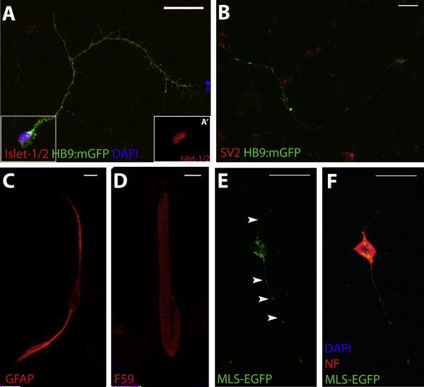
Figure 4. Characterization of primary cultures.
A-D. IHC for various neuronal and non-neuronal markers demonstrate the presence of MNs (Islet-1/2; A, A’(inset of boxed region in A)), synaptic vesicles (SV2; B), glia (GFAP; C) and muscle fibers (F59; D) in primary cultures from 48 hpf HB9:mGFP embryos. E-F. Utilization of MLS-EGFP transgenic zebrafish for primary cultures enables visualization of mitochondria in cell bodies and axons (arrowheads; E) of neurons, as demonstrated by IHC for a neuronal marker (NF; F). GFP expression in transgenic zebrafish lines in MNs is under control of the HB9 promoter (HB9:mGFP; A-D) or in mitochondria using a mitochondrial-localization signal (MLS-EGFP; E-F). Images are acquired using an Olympus BX-51 confocal microscope at 60X magnification. Scale bar equals 20 μm.

