Figure 6.
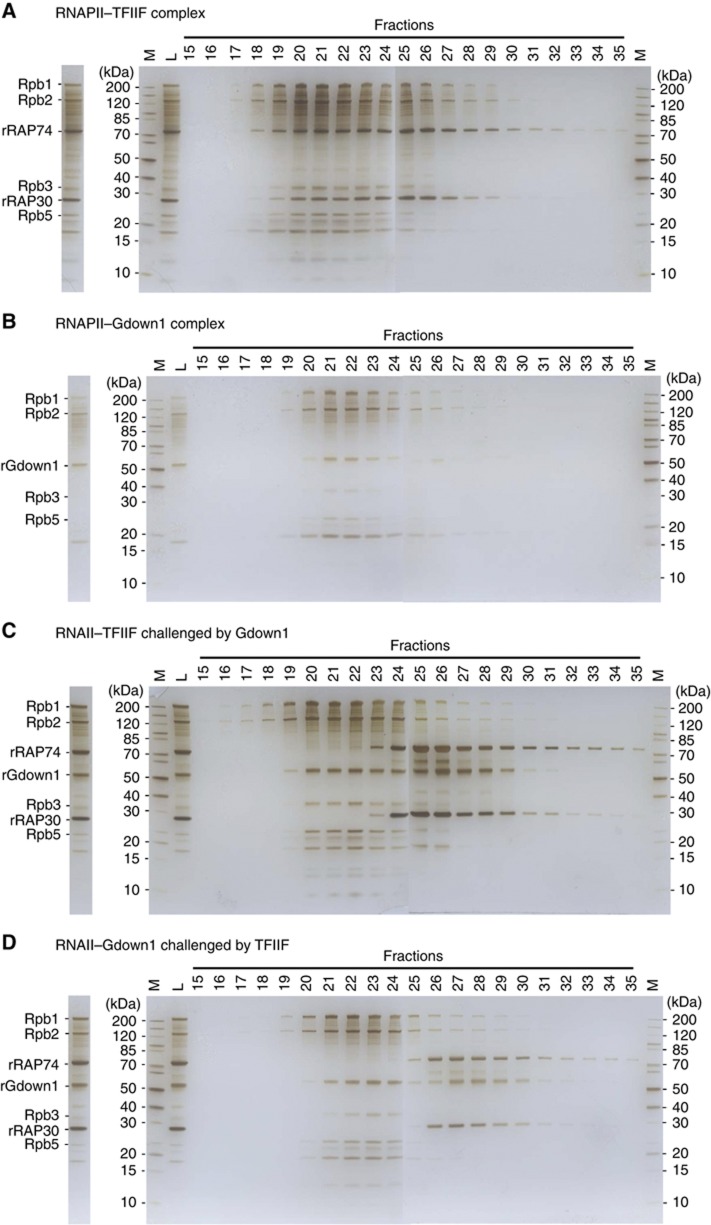
Competition assay using fractionation on a size exclusion column (Superose 6). (A) Major fractions of RNAPII–TFIIF complex (18–22) and major fractions of excess amount of free TFIIF (24–35) are visualized on SDS–PAGE with silver staining. To form RNAPII–TFIIF, eight-fold TFIIF was used. (B) Fractions of RNAPII–Gdown1 complex (20–23) and major fractions of free Gdown1 (26–28). To form RNAPII–Gdown1, four-fold Gdown1 was used (C) The RNAII–TFIIF complex challenged by Gdown1. The RNAPII-associated TFIIF (18–22) now disengages and becomes mostly free (24–35). RNAPII bound TFIIF are replaced by Gdown1 (19–22). (D) The RNAPII–Gdown1 complex challenged by TFIIF. Gdown1 remains mostly associated with RNAPII (21–24) and TFIIF remains free (25–35). Marked bands are major RNAPII subunits: Rpb1 (220 kDa), Rpb2 (133 kDa), Rpb3 (31 kDa), Rpb5 (25 kDa), TFIIF subunits: RAP74 (74 kDa), RAP30 (28 kDa), and rGdown1. In the gel, M stands for marker and L for load. The stained gel strip on the left of each panel represents ∼2.5% of the column load.

