Fig. 5.
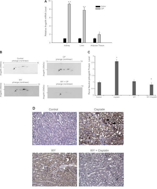
Effect of CP and WY on angiopoietin protein-like 4 (Angptl4) mRNA expression (A), protein levels (B), activity (C), and immunohistochemical localization of Angptl4 (D) in mouse kidney tissues. A: effects of CP on mouse Angptl4 mRNA levels in the kidney, liver, and epididymal white adipose tissues. B: representative autoradiograph of 2-dimensional gel electrophoresis and Western blot analysis from mouse kidney tissue homogenates. The arrows point toward a 35-kDa neutral pI Angptl4 protein in control, CP-, WY-, and WY+CP-treated mice. C: quantitative densitometry of the 35-kDa neutral pI Angplt4 protein shown in B after normalization with β-actin. Values are means ± SE of mRNA levels. Data were obtained from at least 4 independent experiments under each condition. *P < 0.05, **P < 0.001 compared with saline-treated WT mice. †P < 0.05 compared with CP-treated mice in unpaired Student's t-test. D: immunostaining for Angptl4 in kidney tissue. Angptl4 staining is mostly absent in control kidney tissue, and the arrows show that Angptl4 immunostaining is significantly increased in kidney tissue of CP-treated mice, and it is primarily localized to intact proximal tubules (primarily S1 and S2 segments). WY- and WY+CP-treated mice do not show significant staining for Angptl4 in the proximal tubule. Necrotic tubules can be seen only in CP-treated mice (*).

