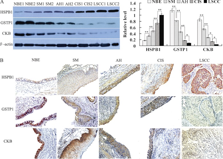
Fig. 2.
Expressional changes of HSPB1, GSTP1, and CKB in the human bronchial epithelial carcinogenic process. A, (left) a representative result of Western blotting shows the expressions of HSPB1, GSTP1, and CKB in the microdissected NBE, SM, AH, CIS, and invasive LSCC; (right) histogram shows the expression levels of the three proteins in these tissues as determined by densitometric analysis. β-actin is used as the internal loading control. Columns, mean from 10 cases of tissues; bars, S.D. (*, p < 0.05; **, p < 0.01 by One-way ANOVA). B, a representative result of immunohistochemistry shows the expression of HSPB1, GSTP1, and CKB in the NBE, SM, AH, CIS, and invasive LSCC. Original magnification, ×200.

