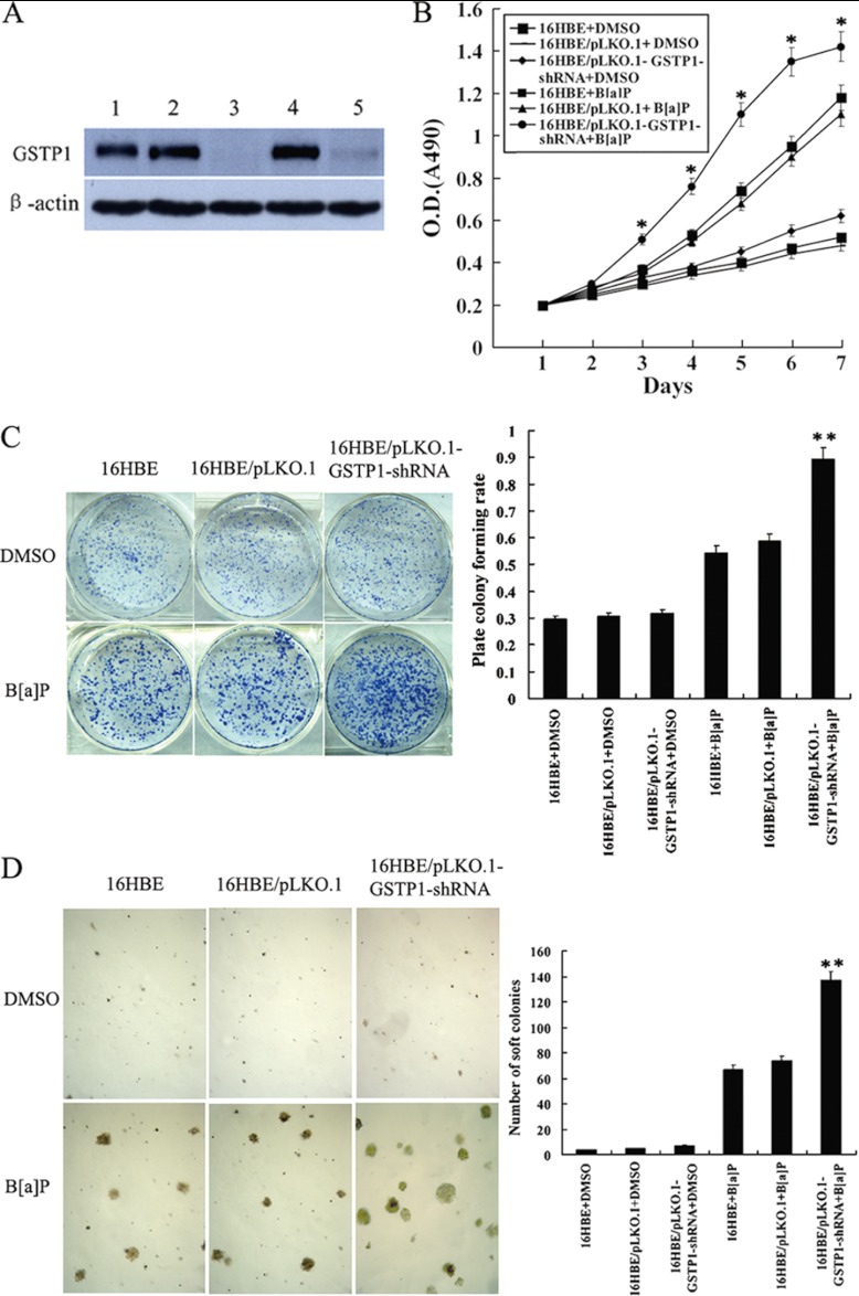
Fig. 4.
The effects of GSTP1 gene knockout on the B[a]P-induced human bronchial epithelial cell transformation. A, Western blotting shows GSTP1 expression in the untransfected (1), empty vector pLKO.1-transfected (2, 4), and pLKO.1-GSTP1-shRNA-tansfected 16HBE cells (3, 5). β-actin is used as an internal control for loading. B, Cell growth in low serum medium after exposed to B[a]P for 16 weeks. Cells were subjected to MTT assay as described in “Experimental Procedures.” Three experiments were done; points, mean; bars, S.D. (*, p < 0.05 versus untransfected or empty vector-transfected 16HBE cells after exposed to B[a]P by Student's t test). C, anchorage dependent colony growth after cells exposed to B[a]P for 16 weeks. (left) cells were subjected to plate colony formation assay as described in “Experimental Procedures,” and colonies were stained with crystal violet and photographed under microscope; (right) the histogram showed plate colony formation rates. Three experiments were done; columns, mean; bars, S.D. (**, p < 0.01 versus untransfected or empty vector -transfected 16HBE cells after exposed to B[a]P by One-way ANOVA). D, Anchorage independent colony growth after cells exposed to B[a]P for 16 weeks. (left) cells were subjected to soft agar colony formation assay as described in “Experimental procedures,” and colonies were photographed under microscope; (right) the histogram showed number of soft agar colonies in 10 randomly chosen microscopic fields using a 5× objective. Three experiments were done; columns, mean; bars, S.D. (**, p < 0.01 versus untransfected or empty vector -transfected 16HBE cells after exposed to B[a]P by One-way ANOVA). Cell proliferation, and plate and soft agar colony growth of the cells exposed to vehicle (DMSO) for 16 weeks are also shown and used as controls. 16HBE, untransfected cells; 16HBE/pLKO.1, empty vector pLKO.1-transfected cells; 16HBE/pLKO.1-GSTP1-shRNA, pLKO.1-GSTP1-shRNA -tansfected cells.

