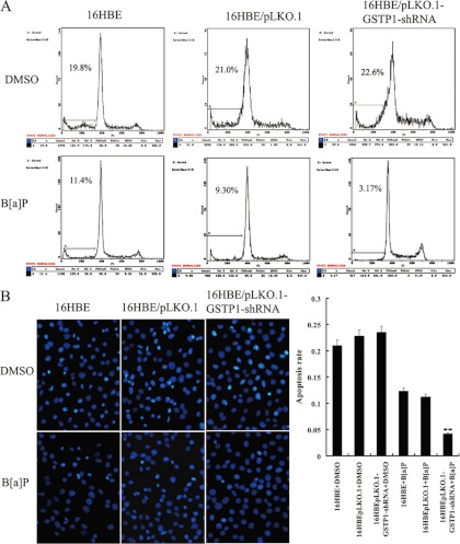
Fig. 5.
The effects of GSTP1 knockdown on the apoptosis of B[a]P-transformed human bronchial epithelial cells. A, A representative result of flow cytometry analysis of cell apoptosis cultured in serum free medium after exposed to B[a]P for 16 weeks. Cells were grown in serum free DMEM medium for 24h, and then assessed for apoptosis by flow cytometry as described in “Experimental procedures.” B, (right) Hoechst 33258 staining of cell apoptosis cultured in serum free medium after exposed to B[a]P for 16 weeks. Cells were grown in serum free DMEM medium for 24h, and then assessed for apoptosis using the cell-permeable DNA dye Hoechst 33258. Apoptotic nuclei showing intense fluorescence corresponding to chromatin condensation; (left) a histogram showed the cell apoptotic rates. Three experiments were done; columns, mean; bars, S.D.(**, p < 0.05 versus untransfected or empty vector -transfected 16HBE cells exposed to B[a]P). Apoptosis of the cells exposed to vehicle (DMSO) for 16 weeks is also shown and used as controls. 16HBE, untransfected cells; 16HBE/pLKO.1, empty vector pLKO.1-transfected cells; 16HBE/pLKO.1-GSTP1-shRNA, pLKO.1- GSTP1-shRNA -tansfected cells.

