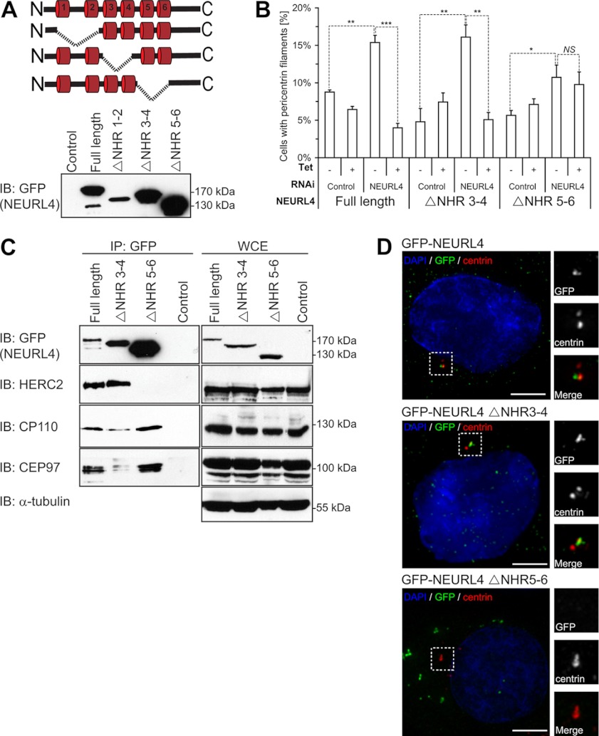
Fig. 4.
Structure-function analysis of NEURL4. A, schematic representation of the different deletion constructs generated for GFP-NEURL4 protein (top panel). Stable U2OS cell lines were generated that expressed each of these deletion constructs, and protein expression was confirmed by immunoblot analysis with the GFP antibody (bottom panel). B, U2OS cell expressing inducible GFP-tagged NEURL4 or NHR deletion mutants were transfected with luciferase esiRNA or esiRNA targeting the 3′-UTR region of NEURL4. Presence of pericentrin filaments was quantified 48 h after induction with 1 μg/ml tetracycline (where indicated) and 72 h after RNAi transfection. The graph shows the averages ± S.E. of the number of cells with pericentrin filaments, obtained in three independent experiments. At least 100 cells were analyzed under each condition. t test results comparing the indicated data sets are shown in the graph. *, p value < 0.05; **, p value < 0.001; ***, p value < 0.0001. NS, nonsignificant difference. C, U2OS stable cell lines expressing the GFP-tagged NEURL4, NEURL4 ΔNHR3–4, and NEURL4 ΔNHR5–6 constructs were induced for expression with 1 μg/ml tetracycline for 16–18 h before harvesting. The exogenously expressed GFP-NEURL4 was immunoprecipitated using GFP antibodies, and the immune complexes were subjected to immunoblot analysis with antibodies against GFP, HERC2, CP110, and CEP97 (left panel). A control IP was performed in parallel in U2OS stable cell lines expressing an empty vector. WCEs were immunoblotted with antibodies to the same proteins as well as the α-tubulin as a loading control (right panel). The control samples were WCEs from U2OS cell line expressing empty vector. HERC2 is a 528-kDa protein and runs as a very high molecular mass band, and there is no molecular mass standard in that region of the gel. D, U2OS cells expressing inducible GFP-tagged NEURL4, NEURL4 ΔNHR3–4, and NEURL4 ΔNHR5–6 were treated with 1 μg/ml tetracycline for 48 h to induce expression. The cells were labeled for DNA (blue; DAPI), pericentrin (red), and GFP (green). Scale bars, 5 μm. GFP-NEURL4 ΔNHR3–4 is co-localized with pericentrin, whereas GFP-NEURL4 ΔNHR5–6 is excluded from the centrosomal region. The insets are magnifications of the outlined regions. The protein size (kDa) is indicated for each immunoblot. DAPI, 4′,6′-diamino-2-phenylindole; IB, immunoblot.

