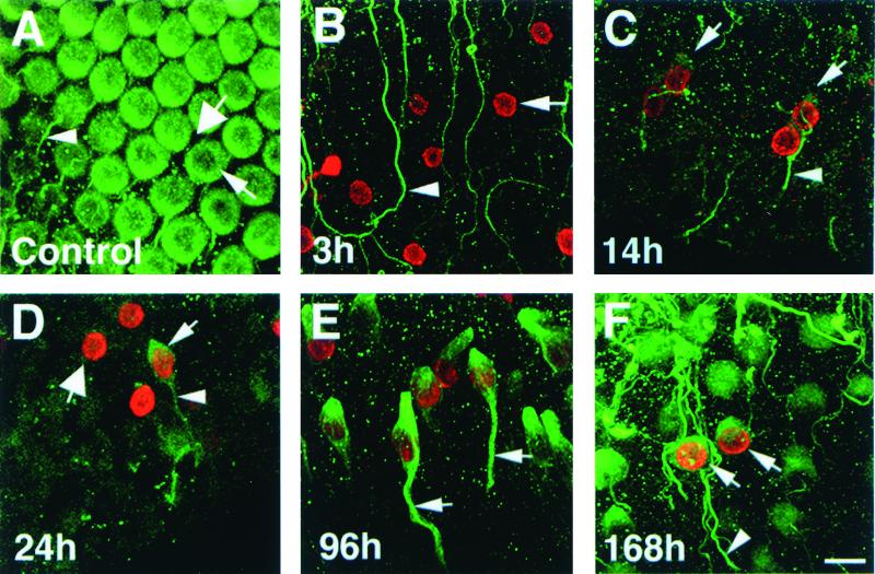
Figure 2.
Temporal progression of hair cell differentiation disclosed by antibodies to BrdUrd and the hair cell-selective protein, class III β-tubulin. Whole-mount preparations of the basilar papilla (BP) labeled with antibodies to BrdUrd (red) and/or β-tubulin (green). (A) In the control BP, β-tubulin is present in hair cells (thin arrow) and nerves (arrowhead), but not supporting cells (thick arrow). (B–F) Drug-damaged BPs taken from chicks at different times after a single BrdUrd injection at 3 days postgentamicin. (B) 3 h postBrdU. Progenitor cells in S or G2 phase of the cell cycle (arrow) are labeled with BrdUrd, but not β-tubulin. β-tubulin is present in nerves (arrowhead) remaining after hair cell loss. (C–F) BrdUrd labeling in regenerated cells at progressively later stages of differentiation. (C) 14 h post-BrdUrd. Some rounded cells (arrows) near the lumen are double-labeled and represent new hair cells at an early stage of differentiation. These cells are associated with nerve processes (arrrowhead). (D) 24 h post-BrdUrd. Regenerated hair cells (thin arrow) are fusiform in shape and are associated with nerve processes (arrowhead). Some BrdUrd-positive cells are not labeled for β-tubulin (thick arrow); these cells are not differentiating as hair cells. (E) 96 h (4 days) post-BrdUrd. Regenerated hair cells have thick cytoplasmic processes that extend toward the lumen and basal lamina (arrows) of the epithelium. (F) 168 h (7 days) post-BrdUrd. Regenerated hair cells (arrows) resemble mature hair cells morphologically. Arrowhead points to nerve process. (Scale bar = 10 μm.)

