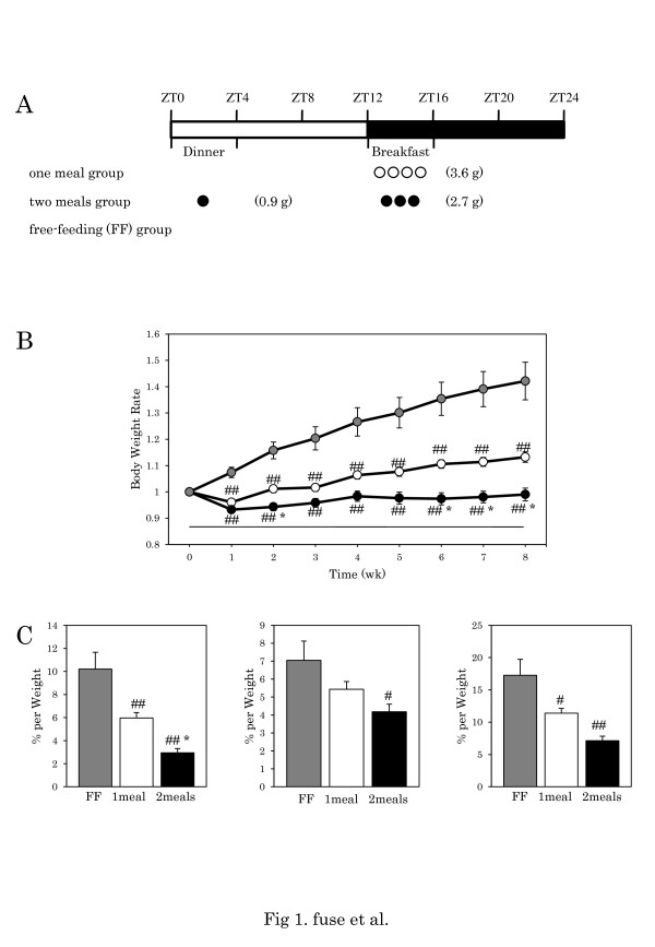
Figure 1.
Body weight gain and quantity of adipose tissue.A: Experimental schedule. Eight-week-old mice were divided into three groups as follows: 3.6 g of high-fat chow at ZT12 as breakfast (one meal group); and 2.7 g of high-fat chow at ZT12 as breakfast and 0.9 g at ZT0 as dinner (two meals group), and a free-feeding (FF) group. White circle: one meal; black circle: two meals. B: Increase in body weight. Body weight at the start of restricted feeding is designated as 1. Restricted feeding starts at 0 (X-axis). Gray circle: FF; white circle: one meal; black circle: two meals. Data are presented as mean ± SEM values (FF, n = 16; one meal, n = 20; two meals, n = 19). # p < 0.05, ## p < 0.01 vs. FF, * p < 0.05, vs. one meal (Tukey-Kramer test). C: Visceral fat, subcutaneous fat and total body fat. Y-axis: (adipose tissue weight/body weight) × 100 (%). Gray column: FF; white column: one meal; black column: two meals. Data are presented as mean ± SEM values (FF, n = 16; one meal, n = 20; two meals, n = 19). # p < 0.05, ## p < 0.01 vs. FF, * p < 0.05, ** p < 0.01 vs. one meal (Tukey-Kramer test).

