Figure 1.
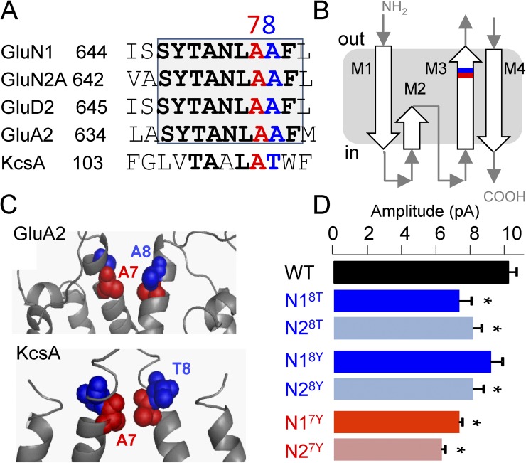
Lurcher-motif residues control NMDA receptor unitary current amplitude. (A) Sequence alignment of the external segment of M3 highlights the conserved lurcher motif; the residues tested here are in red (A7) and blue (A8). This color scheme is used in all subsequent panels. (B) Cartoon of iGluR subunit membrane topology. (C) Partial model of two diagonally situated subunits based on an AMPA receptor (Protein Data Bank accession no. 3KG2) and on KcsA (Protein Data Bank accession no. 1BL8) structures. (D) Unitary current amplitudes of NMDA receptors carrying lurcher (A8T) or lurcher-like (A7Y) substitutions in the N1 or N2A subunits. *, statistically significant differences relative to WT (P < 0.05; one-way ANOVA).

