
Figure 4.
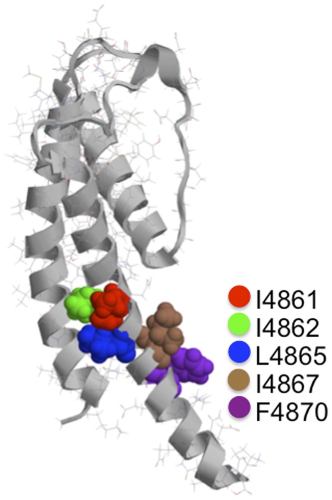
Location of hydrophobic residues in the proposed cytosolic cavity–lining helix (TM10) of RyR2. The figure shows one monomer of the Welch et al. (2004) model of the RyR2 PFR. The luminal entrance to the pore is at the top of the structure, and the cytosolic entrance is at the bottom. The hydrophobic residues of TM10 investigated in this study are highlighted, shown in space-fill, and identified in the key. Molecular graphics were rendered with Ras Top 2.0.2 software.