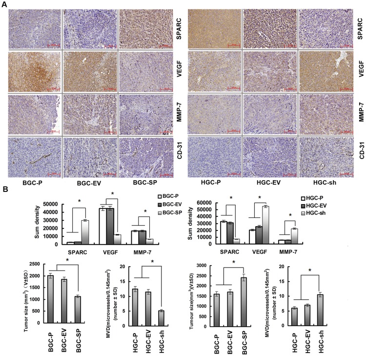
Figure 5. Overexpression of SPARC in gastric cancer cells inhibits tumour development and vascularisation in nude mice.
(A) Paraffin-embedded sections of xenografted tumours were used for immunohistochemical analysis of SPARC, MMP-7, VEGF, and CD-31. (B) Sections were stained with a monoclonal antibody against human SPARC, VEGF, MMP-7. Sum densities were calculated and analyzed by IPP 6.0. Columns are means (±s.d.) of quadruplicate determinations from six mice in each group. *P<0.05, significant difference from control cells. (C) MVD in tumour tissues was quantified by counting CD31-positive areas in each microscopic field of view. Columns are means (±s.d.) of quadruplicate determinations from six mice in each group. *P<0.05, significant difference from control cells. The tumour volume was calculated (volume = width2× length× 0.52). The volume of tumours at the 50th day is indicated by the mean values (±s.d.) of six mice in each group, *P<0.05, significant difference from control cells.

