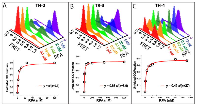
Figure 3.
Single molecule FRET experiments probing GQ unfolding at different RPA concentrations for (A) TH-2; (B) TR-3; and (C) TH-4. When GQ is folded, the donor and acceptor dyes are close to each other, resulting in high-FRET efficiency. RPA binding to DNA induces GQ unfolding, separating the fluorophores away from each other and resulting in low FRET efficiency. As the RPA concentration is increased, the population of the low FRET peak gradually increases as more GQ constructs are unfolded. For TR-3 and TH-4 the fraction of unfolded GQ structures saturates at certain value and complete unfolding of all molecules cannot be attained. The panels at the bottom show a Langmuir isotherm fit to the unfolding data.

