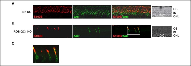
Fig. 10.
Immunohistochemical localization of S100B in the retinas of the Nrl KO and ROS-GC1 KO mice. Cryosections from Nrl KO (A) and ROS-GC1 KO (B) mice retinas were stained with antibodies against S100B and cone arrestin. To focus on the photoreceptors, only the photoreceptor and part of the outer nuclear layers are shown. Nrl KO mice do not develop rods in their retinas; all photoreceptors are cones; in the retinas of ROS-GC1 KO mice, cones degenerate. Images labeled “S100B” and “cArr” show immunostaining with only S100B or cone arrestin antibody. Images labeled “S100B/cArr” show co-staining with both antibodies. The region framed in figure (B) “S100B/cArr” is enlarged as (C). DIC images show integrity of the sections.

