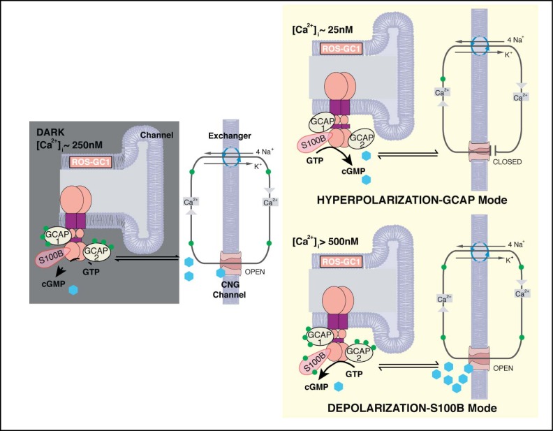
Fig. 12.
Two modes of ROS-GC1 modulation by [Ca2+]i in cones. Dark state (left). At [Ca2+]i = 250 nM Ca2+ sensors: GCAP1, GCAP2, S100B are ROS-GC1 bound; GCAP1 and GCAP2 are [Ca2+]i bound; ROS-GC1 is in the basal state. Cyclic GMP generated keeps a fraction of CNG channels open allowing influx of Na+ and Ca2+. Hyperpolarization-GCAP mode (Top right). LIGHT triggers activation of phosphodiesterase and hydrolysis of cyclic GMP. The decrease in cyclic GMP causes CNG channels to close, preventing influx of Na+ and Ca2+ and hyperpolarizing the outer segment plasma membrane. Extrusion of Ca2+ by the Na+/ Ca2+-K+ exchanger lowers [Ca2+]i from 250 nM to about 25 nM. The decline causes GCAPs to stimulate ROS-GC1. Depolarization-S100B mode (Bottom right). In the pathological state, [Ca2+]i levels rise above 500 nM. Ca2+ is captured by S100B, triggering activation of ROS-GC1. Cyclic GMP formed opens more CNG channels, increasing the influx of Na+ and Ca2+ and raising [Ca2+]i to even higher levels. Extrusion of Ca2+ through Na+/Ca2+-K+ exchanger slows as the ion gradients collapse. The cone outer segment membranes stay in ever-DARK-state.

