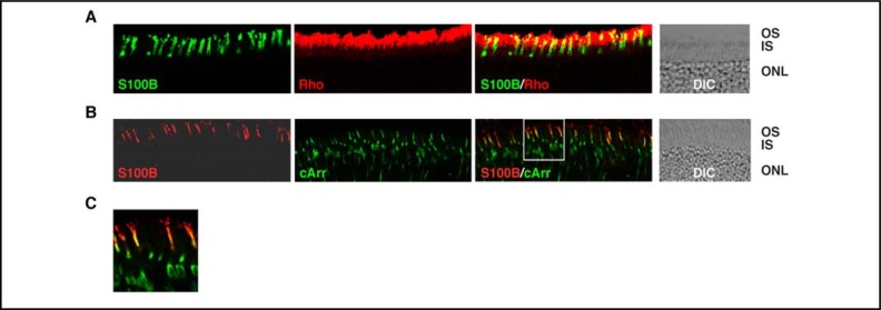
Fig. 9.
Expression of S100B in cones. Cryosections from WT mouse retina were doubly immunostained with antibodies against S100B and rhodopsin (A) or S100B and cone arrestin (B). To focus on the photoreceptors, only the photoreceptor and part of the outer nuclear layers are shown. Images labeled “S100B”, “Rho” (rhodopsin) and “cArr” (cone arrestin) show immunostaining with the respective antibody only; images labeled “S100B/Rho” and “S100B/cArr” show the composites. The region framed in the “S100B/cArr” image is enlarged as (C). For reference, DIC images show the retinal layers of the sections.

