
Fig 4.
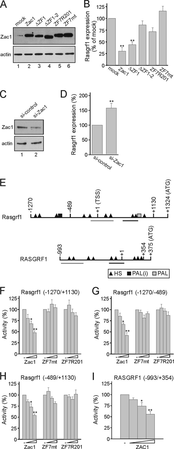
Zac1 inhibits Rasgrf1 gene expression. (A) Immunoblot analysis of Zac1 constructs transfected into predifferentiated R7T1 β cells. WCE (25 μg) were tested with the indicated antibodies. (B) Zac1 represses Rasgrf1 expression in predifferentiated R7T1 β cells. Following the transfection of Zac1 constructs, Rasgrf1 expression was determined by qRT-PCR analysis and normalized to the result from mock transfection, which was set to 100%. (C and D) Knockdown of Zac1 derepresses Rasgrf1. Control or Zac1 siRNA was transfected in predifferentiated R7T1 β cells, which were harvested 2 days later. (C) Immunoblot (50 μg WCE) shows Zac1 expression in control or Zac1 siRNA-transfected R7T1 cells. (D) Derepression of Rasgrf1 was investigated by qRT-PCR analysis; the expression of Rasgrf1 under control siRNA treatment was set to 100%. (E) Scheme of the mouse and human Rasgrf1 promoter. Transcriptional start site (TSS), translation start codon (ATG), and potential Zac1 DNA-binding sites are shown; abbreviations are half-site (HS), imperfect palindrome [PAL(i) (G3C4)], and palindrome [PAL (G5C4)]. In contrast to mice, the human RASGRF1 promoter contains only half-site repressor DNA elements. Highly conserved regions (>85%) between mouse and human Rasgrf1 promoters are underlined. (F to H) Zac1, Zac-ZF7mt, or Zac-ZF7R201 was transfected (100, 250, and 500 ng each) in predifferentiated R7T1 β cells together with the indicated Rasgrf1 promoter reporter constructs (2 μg). (I) Cotransfection of ZAC1 (50, 100, and 250 ng) in predifferentiated R7T1 β cells leads to the repression of a human RASGRF1 promoter reporter construct (2 μg). Data represent means ± SD from three (B and D) or five (F to I) independent experiments performed in duplicate. *, P < 0.05; **, P < 0.01.