Fig 7.

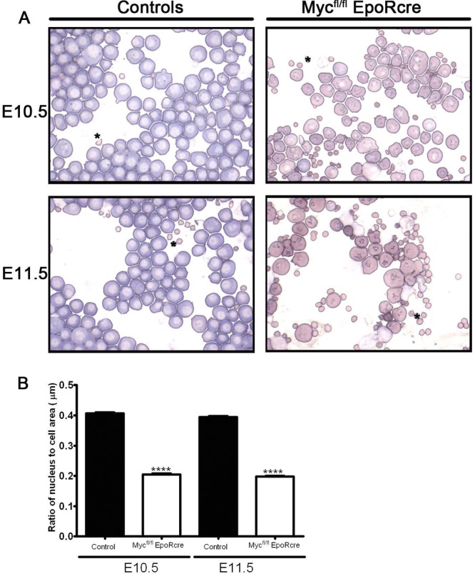
(A) Cytological analysis of circulating primitive erythroid cells. Giemsa-stained cytospin preparations of peripheral blood from E10.5 and E11.5 control (left) and Mycfl/fl; EpoR-Cre (right) embryos. Primitive blood cell preparations of the control embryos at E10.5 and E11.5 exhibited numerous large erythroid cells with features characteristic of basophilic erythroblasts. In contrast, Mycfl/fl; EpoR-Cre embryos had altered Giemsa reactivity and a more orthochromatophilic erythroblast appearance. The Mycfl/fl; EpoR-Cre cells also displayed more condensed and eccentric nuclei in comparison to those of controls, indicating that the Mycfl/fl; EpoR-Cre cells were more mature. Images were obtained with a Zeiss Axiophot microscope. Magnification, ×64. (B) Nuclear-to-cell morphometric analysis indicates a more mature erythroid cell profile in Mycfl/fl; EpoR-Cre embryos. Histograms of ratios of nuclear to cell surface areas of circulating erythroid cells from WT controls (n = 3) and Mycfl/fl; EpoR-Cre embryos (n = 3) at E10.5 and E11.5 were obtained for a minimum of 103 primitive erythroid cells (range, 103 to 203). Importantly, the ratio of nuclear to cell surface areas in Mycfl/fl; EpoR-Cre embryos was more than 2-fold lower than that of controls at E10.5 and E11.5 (****; P < 0.0001). In controls, the ratio of nucleus to cell surface area decreased significantly between E10.5 to E11.5 (P < 0.006).
