Fig 5.
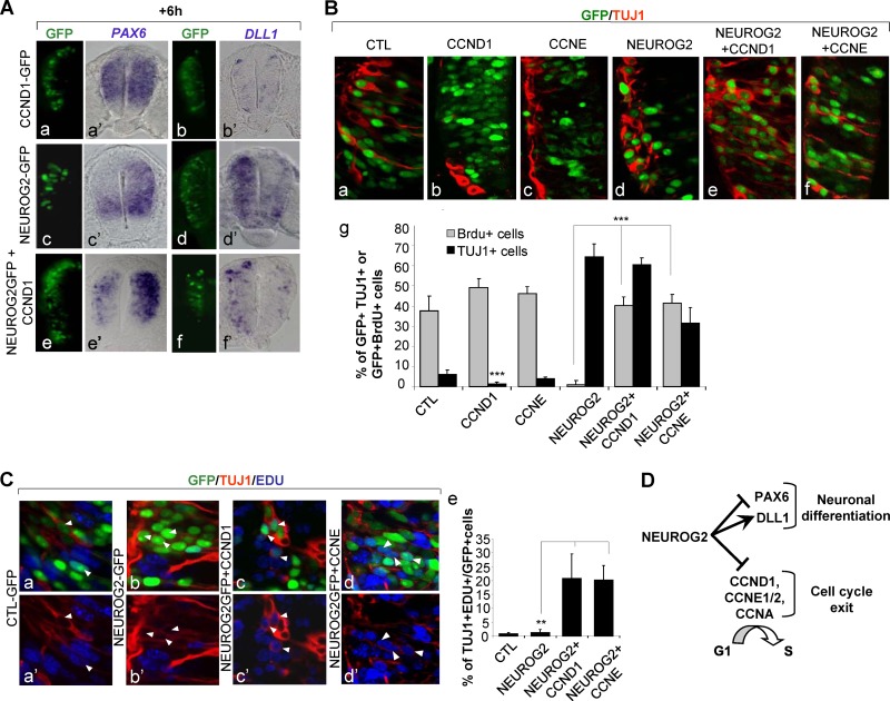
NEUROG2 can control differentiation independently of cell cycle exit. (A) Detection of PAX6 (a′, c′, and e′) and DLL1 (b′, d′, and f′) transcripts on transversal sections by in situ hybridization 6 h following electroporation with CCND1 (a and b′), NEUROG2 (c and d′), or NEUROG2 and CCND1 (e and f′). PAX6 and DLL1 transcripts are modified by NEUROG2 even in the presence of high levels of cyclin D1. (B) Detection of GFP+ TUJ1+ cells 24 h postelectroporation with the mentioned constructs. (a and f) Close-up on transversal sections of embryos electroporated with the mentioned constructs 24 h following electroporation and analysis of TUBB3 (Tuj1). GFP in green; Tuj1 in red. (g) Histograms presenting the T quantification of GFP+ Tuj1+ cells (in black) and GFP+ BrdU+ cells (in gray) in the mentioned experimental conditions, 24 h following electroporation (**, P < 0.01; ***, P < 0.001; Student t test). (C) Detection of GFP+ TUJ1+ EDU+ cells 24 h postelectroporation with the mentioned constructs. (a to d′) Close-up on transversal sections; GFP in green, EdU in blue, TUJ1 in red. (e) Histograms presenting the quantification of TUJ1+ EDU+ cells among the GFP+ cells in the mentioned experimental conditions (**, P < 0.01; ***, P < 0.001; Student t test). (D) NEUROG2 can control neuronal differentiation independently of cell cycle exit.

