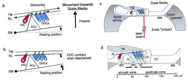
Figure 1.
(a) A model of hair cell excitation without OHC motility according to Davis (17) (see text for details). (b) OHCs contract when depolarized and the BM is drawn to the reticular lamina. (c) Experimental arrangement with laser diode beam. (d) Transverse section through the organ of Corti in the 15.5-kHz region based on measurements made in vivo and from histological sections: scale bar referenced to the bony edge of the spiral lamina. Solid horizontal bars indicate the following regions across the BM width with respect to the bony edge of the spiral lamina: 30–50 μm (junction of inner and outer pillar cells), 60–70 μm (near midpoint of OPC base), 80–120 μm (Deiters' cells). TM, tectorial membrane; IHC, inner hair cell; OHC, outer hair cell; HC, Hensen cell; CC, Claudius cell region; PCs, pillar cells; IPC, inner pillar cell; OPC, outer pillar cell; DC, Deiters' cell; RL, reticular lamina; TC, tunnel of Corti. Experiments were performed on deeply anaesthetized pigmented guinea pigs (180–300 g, 0.06 mg atropine sulfate s.c., 30 mg/kg pentobarbitone i.p., 4 mg/kg Droperidol i.m.; 1 mg/kg Phenoperidine i.m.), which were tracheotomized, artificially respired, and with core temperatures maintained at 37°C. Modified from Nilsen and Russell (22).

