
Fig 4.
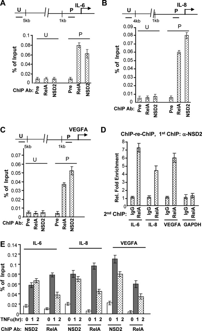
NSD2 associates with NF-κB at endogenous target gene promoters. (A to C) PC-3 cells were growing in regular growth medium and harvested for ChIP assays with preimmune serum or anti-NSD2 or anti-RelA/p65 antibodies (Ab). ChIP and input DNA was analyzed by real-time PCR with primers amplifying the indicated genomic regions of individual genes (U, upstream; P, promoter). ChIP data obtained from triplicate experiments were presented as a percentage of input signals. (D) The first ChIP was performed as described for panels A to C with NSD2 antibody. The immunoprecipitation complex was eluted and subject to the second ChIP procedures with control IgG or anti-RelA antibody. DNA from the second ChIP was analyzed by real-time PCR, as described for panels A to C. Data from control IgG were set as 1. (E) PC-3 cells were serum deprived for 16 h (to diminish NF-κB activation by undefined factors) and then treated with 10 nM TNF-α for the indicated times (hours) and harvested for ChIP assays with anti-NSD2 or anti-RelA/p65 antibodies, as described for panels A to C.