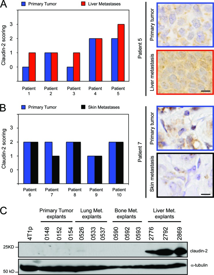
Fig 1.
Claudin-2 expression is enriched in human breast cancer liver metastases compared with matched primary breast tumors. (A and B) Paraffin-embedded sections from five matched primary breast tumors and liver metastases (A) and five matched primary breast tumors and skin metastases (B) were subjected to immunohistochemical staining with an anti-claudin-2 antibody. The scoring of claudin-2 staining was performed by two independent reviewers. (A) Claudin-2 staining was enriched in three of five liver metastases compared to the matched primary breast tumors. The scale bar (bottom) represents 10 μm. (B) No enrichment in claudin-2 staining was observed for the skin metastases compared to the matched primary breast tumors. The scale bar (bottom) represents 10 μm. (C) Immunoblot analysis of claudin-2 demonstrates expression specifically in 4T1-derived explant populations isolated from liver metastases compared to those derived from lung or bone metastases. As a loading control, total cell lysates were blotted for α-tubulin.

