Fig. 6.
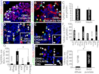
Overexpression of Cyclin D3 and Cdk6 activates the DNA damage response in human β-cells. Dual immunostaining for γH2AX (green), DAPI (blue), and (A) Pdx1 (red), (B) insulin (red), (D) BrdU (red), and (E) Ki67 (red) in Cyclin D3 and Cdk6-transduced human islets. C, Quantification of the percentage of either Pdx1+, or insulin+ cells that are γH2AX+ in primary human islets transduced with both Cdk6 and Cyclin D3 (n = 3–4 per group). F, Quantification of the percentage of either BrdU+,total, BrdU+,diffuse, BrdU+,punctate, Ki67+,total, Ki67+,normal, or Ki67+,irregular cells that are γH2AX+ in primary human islets transduced with both Cdk6 and Cyclin D3 (n = 3–4 per group). G, Quantification of the percentage of γH2AX+ cells that are either BrdU+,diffuse, BrdU+,punctate, Ki67+,normal, or Ki67+, irregular (n = 3–4 per group; *, P < 0.001 BrdU+,punctate vs. Ki67+,irregular populations). H, Immunostaining for phospho-p53 on serine 15 (green), DAPI (blue), and BrdU (red) in Cyclin D3 and Cdk6-transduced human islets. I, Quantification of the percentage of either BrdU+,diffuse or BrdU+,punctate that are phospho-p53 (serine 15)+ (n = 4; *, P < 0.001). The white arrowheads indicate colocalization, and yellow arrowheads indicate noncolocalization between two markers. All human islets were incubated for 72 h after transduction. The scale bars indicate 25 μm in panels A and H and 5 μm in the insets of panels E and H. p, Punctuate; d, diffuse; n, normal; i, irregular.

