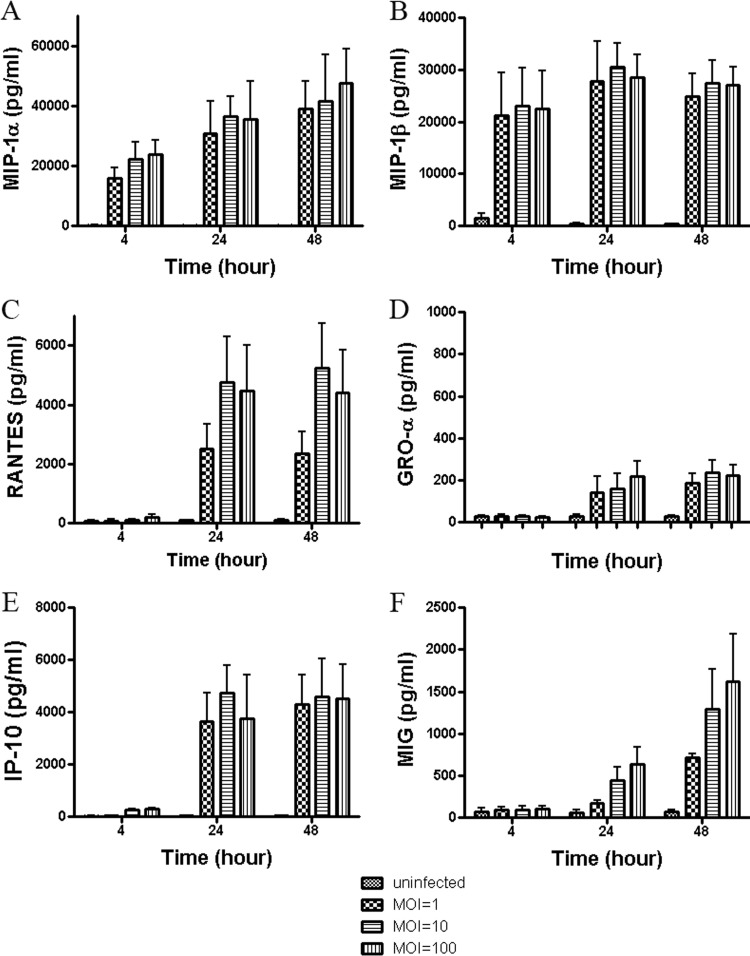
Fig 1.
Chemokine induction in human DCs infected with C. jejuni 81-176. Culture supernatants were tested at 4, 24, and 48 h postinfection with C. jejuni 81-176 at various MOIs of 1, 10, or 100. Phosphate-buffered saline-incubated DCs served as uninfected controls. Chemokine assays were performed by using human chemokine bead kits and a Luminex 100 analyzer. Data analysis was performed using MasterPlex QT quantitation software (MiraiBio, Alameda, CA). The levels of MIP-1α, MIP-1β, RANTES, GRO-α, IP-10, and MIG are shown in A, B, C, D, E, and F, respectively. Data are presented as the means ± SD of duplicate wells from at least 3 independent assays, and ANOVA was used for statistical comparisons among three groups.

