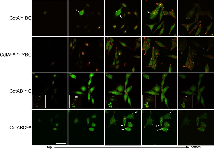
Fig 3.
Live-cell imaging of CHO-K1 cells exposed to 10 μg/ml of the heterotrimer CdtALumBC, CdtABLumC, or CdtABCLum. Successive Z sections from representative fields were taken from top to bottom. Cells were colabeled with Lumio green (green fluorescence) and WGA-Alexa Fluor 555 (red fluorescence) at 18 h postintoxication. Cells treated with a heterotrimer containing a binding-deficient CdtA subunit (CdtALum, Y214ABC) were used as a control. The insets in panel CdtABLumC show a representative field of cells containing fragmented nuclei (FN). S, cell surface; P, polar end. Scale bar = 50 μm.

