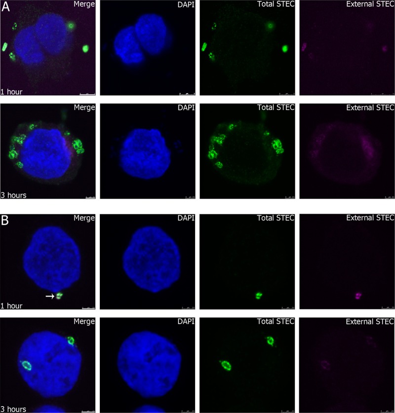
Fig 2.
Association of STEC strains 98NK2 and 98NK2ΔfliC with HCT-8 cells. HCT-8 cells were infected with STEC strain 98NK2 (A) or 98NK2ΔfliC (B) for 1 or 3 h in 8-well chamber slides. Slides were fixed and processed for inside-out immunofluorescence, as described in Materials and Methods. Nuclei are blue (DAPI), extracellular STEC is purple (Alexa Fluor 647), and total STEC is green (Alexa Fluor 488). Bars, 5 μm (top) and 2.5 μm (bottom) (A) and 2.5 μm (B). The white arrow in the merged image in panel B shows colocalized fluorescence indicative of external STEC.

