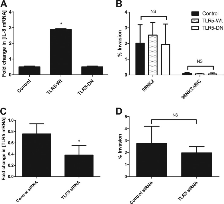
Fig 3.
TLR5 is not involved in invasion of cells by STEC strain 98NK2 or 98NK2ΔfliC. (A) Untransfected HCT-8 cells or cells stably transfected with TLR5-Wt, TLR5-DN, or empty vector (control) were stimulated with strain 98NK2, and RNA was extracted after 4 h. IL-8 mRNA was quantitated by real-time RT-PCR. Results are expressed as the fold increase in the concentration of IL-8 mRNA relative to that in untransfected HCT-8 cells, and data are shown as the means plus SDs for triplicate assays. *, P = 0.0257 compared to control (empty vector) cells or P = 0.0265 compared to cells transfected with TLR5-DN. (B) Invasion assays with STEC strains 98NK2 and 98NK2ΔfliC in HCT-8 cells stably transfected with TLR5-Wt, TLR5-DN, or empty vector. Data shown are the means plus SDs from three independent experiments, each performed in at least duplicate assays (n = 8). NS, not significant. (C) HCT-8 cells were transfected with siRNA directed against TLR5 or with negative-control siRNA, and RNA was extracted 24 h later (as described in Materials and Methods). TLR5 mRNA was quantitated by real-time RT-PCR. Results are expressed as the fold increase in the concentration of TLR5 mRNA relative to control cells. Data shown are the means plus SDs from three independent experiments performed in triplicate. *, P = 0.0106. (D) HCT-8 cells were transfected with siRNA directed against TLR5 or with negative-control siRNA and used in invasion assays performed with strain 98NK2 approximately 48 h later (as described in Materials and Methods). Results shown are the means plus SDs from three independent experiments, each performed in quadruplicate. NS, not significant.

