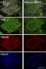
Fig. 2.
Confocal images of abducens motor neurons after normal conditioning for 2 pairing sessions (C2) or conditioning following treatment with the glutamate receptor A4 (GluA4) subunit small-interfering RNA (tGluA4 siRNA). Images are unprocessed except that the contrast was increased for the illustration. After drawing the outline of the cell of interest by the investigator, the software breaks the original image into its individual color channels, revealing punctate staining for synaptophysin (Syn; green), GluA4 (red), or GluA1 (blue) for quantitative analysis. Original scale bar = 10 μm.

