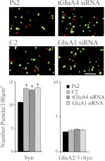
Fig. 9.
Synaptic localization of tGluA2/3-containing AMPARs after conditioning or treatment with the siRNAs. Levels of synaptophysin punctate staining increased significantly in all of the groups that received paired stimulation compared with Ps2 (P < 0.0001). Conditioning in normal medium or after treatment with either the tGluA4 or GluA1 siRNA did not alter the colocalization of these AMPARs with synaptophysin. Representative images of abducens motor neurons are also shown (synaptophysin, green; tGluA2/3, red; colocalization, yellow). *Significant differences from Ps2; scale bar = 2 μm.

