Fig. 3.
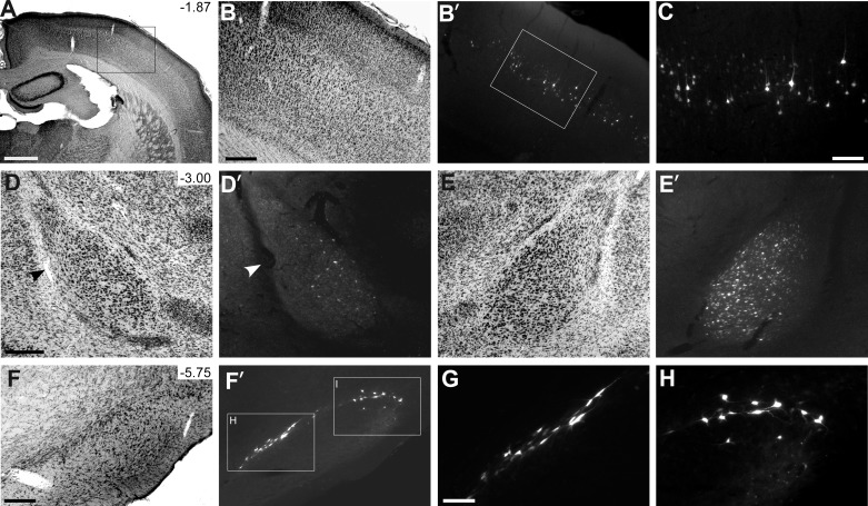
Retrogradely labeled neurons produced by the FG injection shown in Fig. 2. A: section of SI cortex illustrating the region magnified in subsequent panels. B, B': adjacent sections showing the primary somatosensory (SI) cortex and the location of FG-labeled corticostriatal neurons in layer Va. Box indicates the region in C. D, E': bilateral sections showing retrogradely labeled neurons in the basolateral amygdala. F, G': cytoarchitecture of substantia nigra and FG-labeled neurons in the dorsal and lateral subnuclei of the pars compacta. Arrowheads indicate common blood vessels. Scale bars: 1 mm in A; 250 μm in B, D, and F; 100 μm in C and G.

